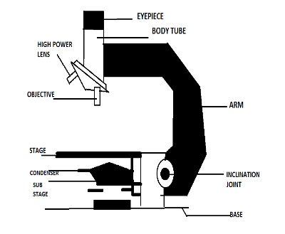
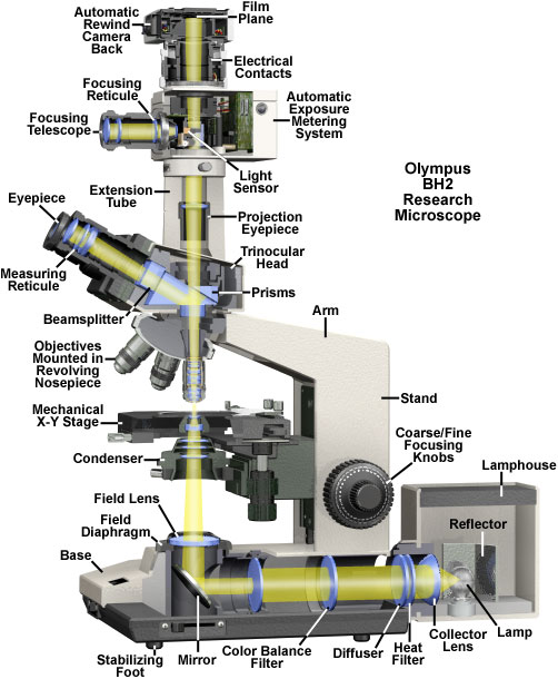
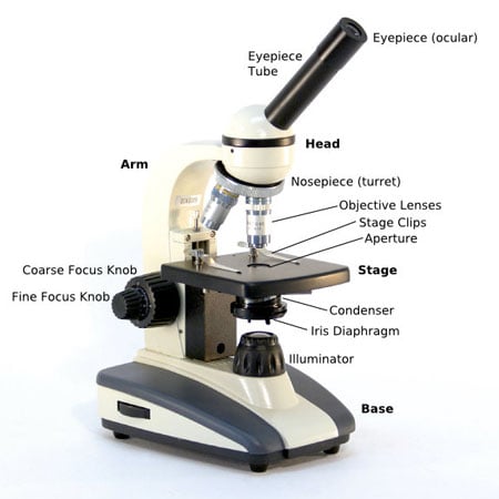
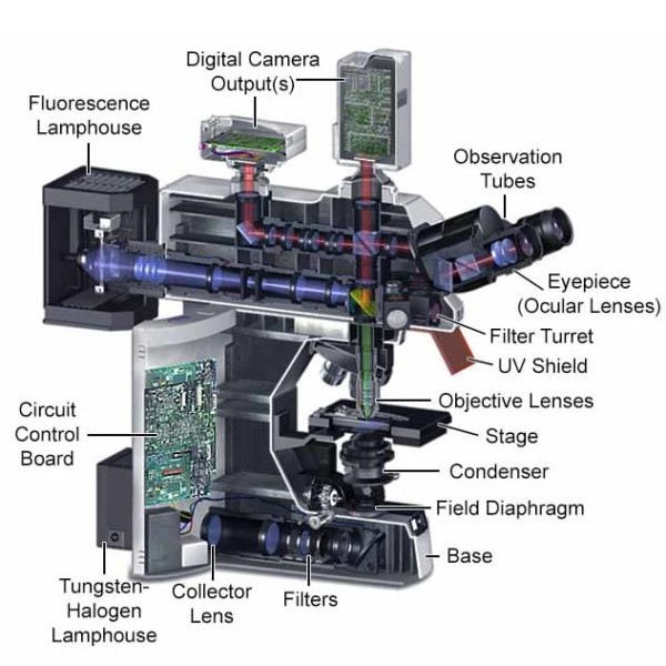
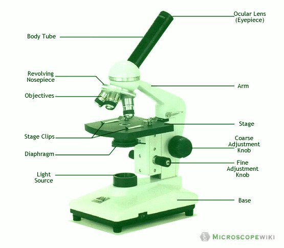
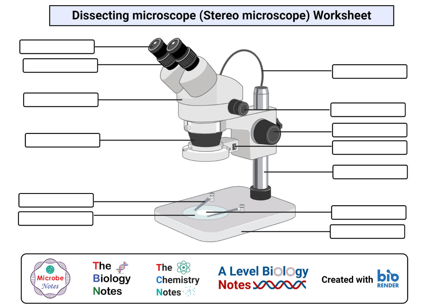
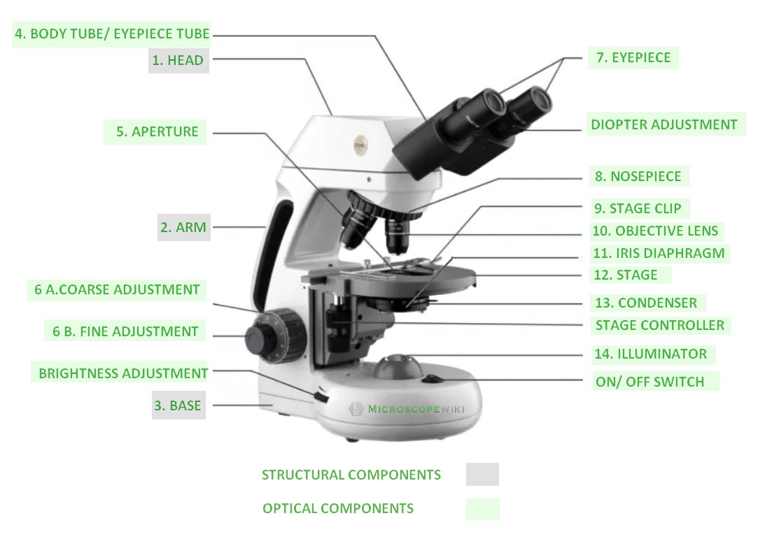
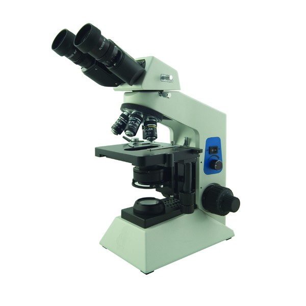
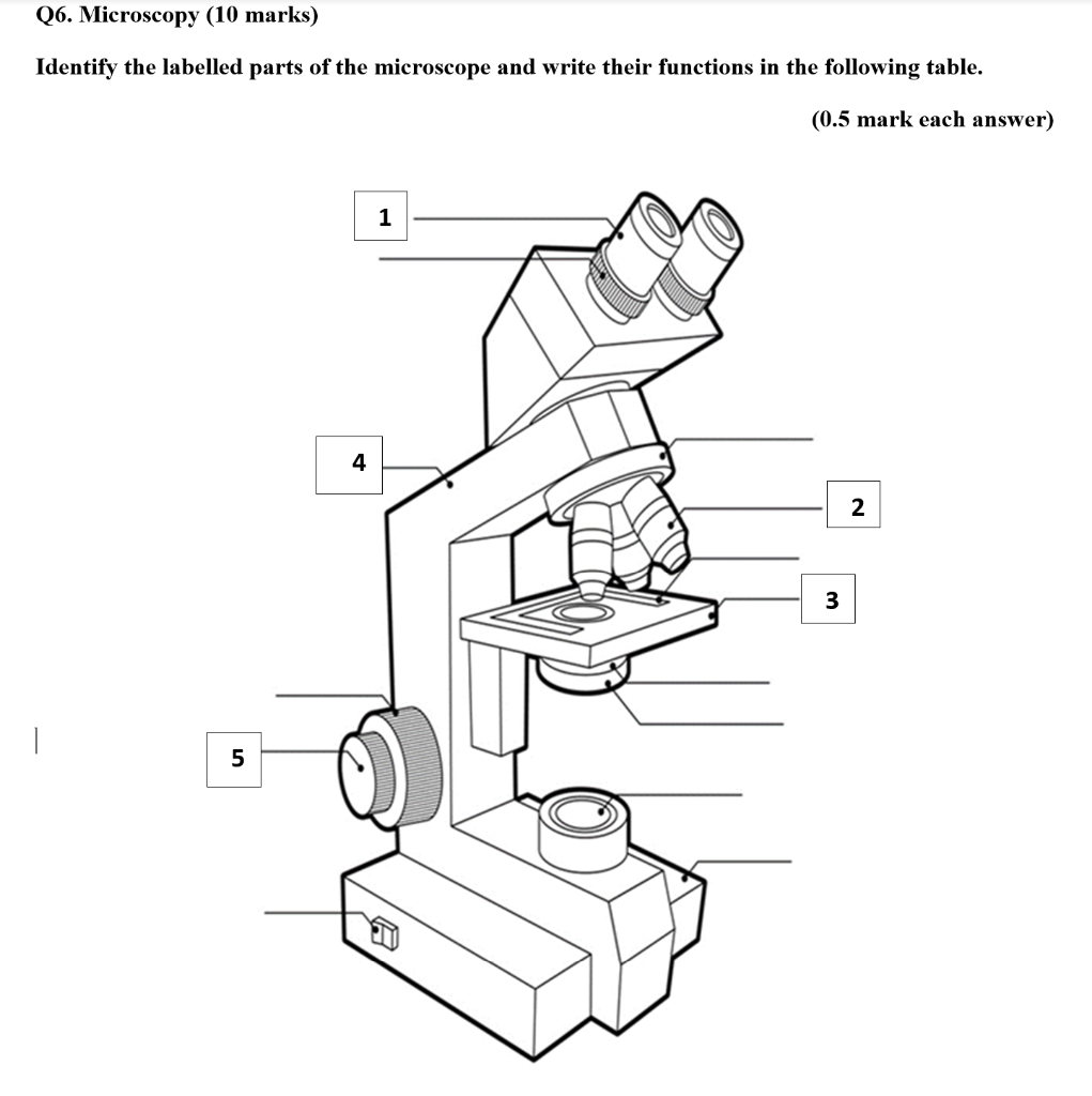
39 labelled diagram of a binocular microscope
Microscope Parts, Types & Diagram | What is a Microscope? Microscope Diagram There are many illustrations available for the diagram of a light microscope. The essential parts include the head, base, arms, lenses, and lights. In diagrams, one... How to Label a Binocular Microscope | Sciencing Label the coarse adjustment knob and the fine adjustment knob. Identify the light source at the bottom of the microscope. The light source is an electrically powered lamp that provides the light used to view the specimen. Many binocular microscopes are equipped with a lamp rheostat that allows the intensity of the light to be adjusted.
Compound Microscope: Definition, Diagram, Parts, Uses, Working ... - BYJUS The field of view of a microscope is defined as how large the area is seen within the eyepiece. The lower the magnification, the smaller the image seen in the eyepiece. As the magnification increases, the size of the image gets bigger although the size of the hole in the eyepiece remains unchanged. What is the depth of field of a microscope?

Labelled diagram of a binocular microscope
What Are the Parts and Functions of a Binocular Microscope? The parts of a binocular microscope are the eye piece (ocular), mechanical stage, nose piece, objective lenses, condenser, lamp, microscope tube and prisms. Each part plays an important role in the microscope's function. Compound Microscope Parts - Labeled Diagram and their Functions Labeled diagram of a compound microscope Major structural parts of a compound microscope There are three major structural parts of a compound microscope. The head includes the upper part of the microscope, which houses the most critical optical components, and the eyepiece tube of the microscope. Parts of the Microscope with Labeling (also Free Printouts) 5. Knobs (fine and coarse) By adjusting the knob, you can adjust the focus of the microscope. The majority of the microscope models today have the knobs mounted on the same part of the device. Image 5: The circled parts of the microscope are the fine and coarse adjustment knobs. Picture Source: bp.blogspot.com.
Labelled diagram of a binocular microscope. What is a Binocular Microscope? (with pictures) - All the Science Binocular microscopes are commonly featured in labs. There are three basic primary types of microscopes: the student, the benchtop and the research microscope. Any of these can be, and probably will be, a binocular microscope. The cheapest of these is the student microscope, which is so named because it is most commonly seen in the classroom. Binocular Microscope Parts Quiz - PurposeGames.com This online quiz is called Binocular Microscope Parts. It was created by member angeline0127 and has 16 questions. ... Label the 6 layers & 4 features of the Sun. Science. English. Creator. teacherrojas. Quiz Type. Image Quiz. Value. 10 points. Likes. 11. Played. 28,860 times. Printable Worksheet. Play Now. Labeling the Parts of the Microscope | Microscope World Resources Labeling the Parts of the Microscope This activity has been designed for use in homes and schools. Each microscope layout (both blank and the version with answers) are available as PDF downloads. You can view a more in-depth review of each part of the microscope here. Download the Label the Parts of the Microscope PDF printable version here. Microscope Labeling Diagram | Quizlet Definition Separates the eyepiece lens from the objective lenses. Location Term Eyepiece Definition Contains the ocular lens and magnifies the image produced by the objective lenses. Location Term Coarse Focus Knob Definition Moves the stage large distances to roughly focus the image. Location Term Fine Focus Knob Definition
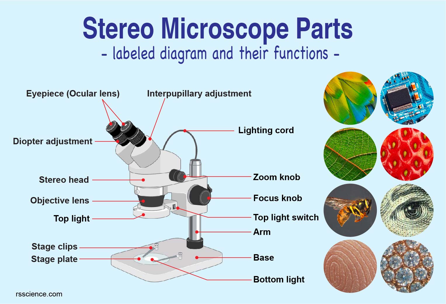
Compound Microscope Parts, Functions, and Labeled Diagram Compound Microscope Definitions for Labels Eyepiece (ocular lens) with or without Pointer: The part that is looked through at the top of the compound microscope. Eyepieces typically have a magnification between 5x & 30x. Monocular or Binocular Head: Structural support that holds & connects the eyepieces to the objective lenses. What Are The Different Parts of a Binocular? - The Ultimate Guide!! So, binoculars consist of a two-barrel chamber, eyepiece lenses, objective lenses, Prisms, diopter knob, and focusing wheel. The objective lens collects the light from the objects, the eyepiece presents the magnified object to the eye and prisms re-invert the flipped images and lengthens the light. Parts of a microscope with functions and labeled diagram - Microbe Notes Figure: Diagram of parts of a microscope There are three structural parts of the microscope i.e. head, base, and arm. Head - This is also known as the body. It carries the optical parts in the upper part of the microscope. Base - It acts as microscopes support. It also carries microscopic illuminators. Parts of Stereo Microscope (Dissecting microscope) - labeled diagram ... Labeled part diagram of a stereo microscope Major structural parts of a stereo microscope There are three major structural parts of a stereo microscope. The viewing Head includes the upper part of the microscope, which houses the most critical optical components, including the eyepiece, objective lens, and light source of the microscope.
16 Essential Microscope Parts: Names, Functions & Labeled Diagram Condenser. The condenser is to focus the light, which passes from the microscopic illuminator to the specimen. This condenser is located just below the diaphragm. This diaphragm is one of the essential parts of the compound microscope, which will help to get an accurate and sharp image. The condenser has a magnification power of 400X and above. Compound Microscope - Diagram (Parts labelled), Principle and Uses What are the 13 parts of a microscope? 1. Eyepiece 2. Eyepiece Tube 3. Objective Lens 4. Stage 5. Stage Clips 6. Nosepiece 7. Fine and Coarse Focus knobs 8. Illuminator 9. Aperture 10. Iris Diaphragm 11. Condenser 12. Condenser Focus Knob 13. The Rack stop Q 5. What are the 11 parts of a compound microscope? Dissecting microscope (Stereo or stereoscopic microscope)- Definition ... Stereo Zoom Dissecting Microscope-They are trinocular or binocular dissecting microscopes with a zooming range of 6.7x-45x. They can be attached to the digital camera which takes photos of the viewing images. ... Parts of a microscope with functions and labeled diagram; Light Microscope- Definition, Principle, Types, Parts, Labeled Diagram ... Microscope Parts and Functions Microscope Parts and Functions With Labeled Diagram and Functions How does a Compound Microscope Work? Before exploring microscope parts and functions, you should probably understand that the compound light microscope is more complicated than just a microscope with more than one lens.
A Study of the Microscope and its Functions With a Labeled Diagram ... To better understand the structure and function of a microscope, we need to take a look at the labeled microscope diagrams of the compound and electron microscope. These diagrams clearly explain the functioning of the microscopes along with their respective parts. Man's curiosity has led to great inventions. The microscope is one of them.
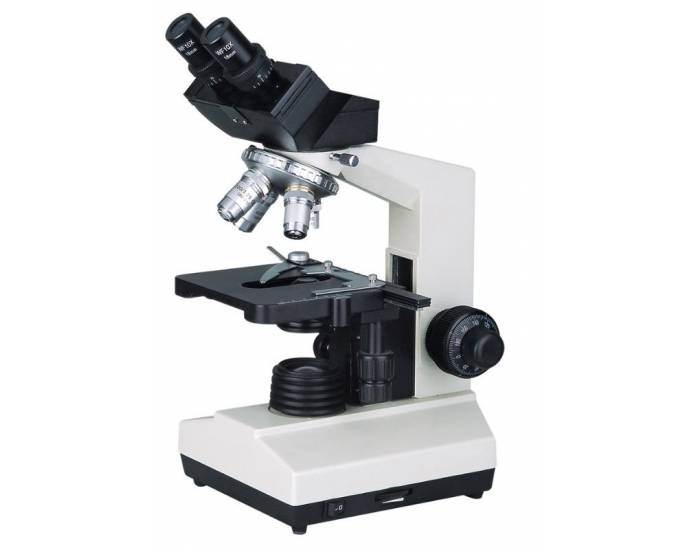
Binocular Microscope Anatomy - Parts and Functions with a Labeled Diagram Now, I will discuss the details anatomy of the light compound microscope with the labeled diagram. Why it is called binocular: because it has two ocular lenses or an eyepiece on the head that attaches to the objective lens, this ocular lens magnifies the image produced by the objective lens. Binocular microscope parts and functions
Parts of a Microscope with Their Functions • Microbe Online The common light microscope used in the laboratory is called a compound microscope. It is because it contains two types of lenses; ocular and objective. The ocular lens is the lens close to the eye, and the objective lens is the lens close to the object. These lenses work together to magnify the image of an object.
PaRTS of a BINOCULAR MICROSCOPE Flashcards | Quizlet one hand should be under this part when microscope is carried substage light provides light source which is projected up through the tissue to allow for viewing condenser adjustment knob raises and lowers the condenser iris diaphragm controls the amount of light passing through the specimen stage Supports the slide and viewing object
Label the microscope — Science Learning Hub In this interactive, you can label the different parts of a microscope. Use this with the Microscope parts activity to help students identify and label the main parts of a microscope and then describe their functions. Drag and drop the text labels onto the microscope diagram.
Microscope: Parts Of A Microscope With Functions And Labeled Diagram. List down the 18 parts of a Microscope. Ocular Lens (Eye Piece) Diopter Adjustment Head Nose Piece Objective Lens Arm (Carrying Handle) Mechanical Stage Stage Clip Aperture Diaphragm Condenser Coarse Adjustment Fine Adjustment Illuminator (Light Source) Stage Controls Base Brightness Adjustment Light Switch
Microscope, Microscope Parts, Labeled Diagram, and Functions Diaphragm or Iris: Most of the laboratory microscopes have a rotating disk under the stage known as diaphragm or iris. Iris Diaphragm controls the amount of light reaching the specimen. The Iris Diaphragm is located above the condenser lens and below the microscope stage.
Types of Microscopes: Definition, Working Principle, Diagram ... - BYJUS The magnifying power of the compound microscope is given as: m = D f o × L f e Where, D is the least distance of distinct vision L is the length of the microscope tube f o is the focal length of the objective lens f e is the focal length of the eyepiece Compound Microscope Diagram Principle of Compound Microscope
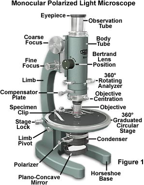
Diagram and labels of a monocular microscope? - Answers A monocular microscope has only one eyepiece while a binocular microscope has two eyepieces with different lenses. Binocular microscopes are more popular today than the monocular microscope for ...
Microscopy: Intro to microscopes & how they work (article) - Khan Academy Magnification is a measure of how much larger a microscope (or set of lenses within a microscope) causes an object to appear. For instance, the light microscopes typically used in high schools and colleges magnify up to about 400 times actual size. So, something that was 1 mm wide in real life would be 400 mm wide in the microscope image.
Sperm Under Microscope with Labeled Diagram - AnatomyLearner These cells show an expanded head, a narrow neck, and an elongated thin (not seen clearly) tail under the microscope. The sample tissue also shows the other spermatogenic cells (primary spermatocytes, secondary spermatocytes, and spermatid) along with the spermatozoa.
Parts of the Microscope with Labeling (also Free Printouts) 5. Knobs (fine and coarse) By adjusting the knob, you can adjust the focus of the microscope. The majority of the microscope models today have the knobs mounted on the same part of the device. Image 5: The circled parts of the microscope are the fine and coarse adjustment knobs. Picture Source: bp.blogspot.com.
Compound Microscope Parts - Labeled Diagram and their Functions Labeled diagram of a compound microscope Major structural parts of a compound microscope There are three major structural parts of a compound microscope. The head includes the upper part of the microscope, which houses the most critical optical components, and the eyepiece tube of the microscope.
What Are the Parts and Functions of a Binocular Microscope? The parts of a binocular microscope are the eye piece (ocular), mechanical stage, nose piece, objective lenses, condenser, lamp, microscope tube and prisms. Each part plays an important role in the microscope's function.










Komentar
Posting Komentar